
Skip to content
The V Foundation logo
Donate
  • About Us

    About the V Foundation

    • Our Mission
    • Financial Responsibility
    • Newsroom

    Team

    • Leadership
    • Partners
    • Ambassadors
    • Careers


  • Research & Impact

    Mission

    • Research
    • Key Research Initiatives
    • Proven Positive Impact

    Granting

    • Award Programs
    • Grant Process
    • Funded Grants

    Resources

    • For Researchers
    • For Patients
  • Take Action

    Donate

    • Ways to Give
    • Become a Monthly Donor
    • Become a Champion
    • Honors & Memorial Giving

    Fundraise

    • DIY Fundraising
    • Run for Team V
    • Cause Marketing


  • Events

    Wine Celebration

    View event

    Chicago Epicurean

    View event

    Capital Epicurean

    View event

    Austin Epicurean

    View event

    Boo-Yah

    View event

    View All Events

  • Donate
  • About Us

    About the V Foundation

    • Our Mission
    • Financial Responsibility
    • Newsroom

    Team

    • Leadership
    • Partners
    • Ambassadors
    • Careers


  • Research & Impact

    Mission

    • Research
    • Key Research Initiatives
    • Proven Positive Impact

    Granting

    • Award Programs
    • Grant Process
    • Funded Grants

    Resources

    • For Researchers
    • For Patients
  • Take Action

    Donate

    • Ways to Give
    • Become a Monthly Donor
    • Become a Champion
    • Honors & Memorial Giving

    Fundraise

    • DIY Fundraising
    • Run for Team V
    • Cause Marketing


  • Events

    Wine Celebration

    View event

    Chicago Epicurean

    View event

    Capital Epicurean

    View event

    Austin Epicurean

    View event

    Boo-Yah

    View event

    View All Events

The Latest

“I’m a survivor, and you can be one too”

Read more
">

Thank you, C2C4C United States Riders!

Read more
">

Dr. Maria Figueroa: The Importance of Teamwork in Science

Read more
">

Andrew Jones – Life After Cancer & Fatherhood

Read more
">
View All

About the Foundation

  • About Us
  • Our Story
  • Leadership Team
  • Partners
  • Financial Responsibility
  • Diversity, Equity and Inclusion
  • Cookie Policy

How to Help

  • Ways to Give
  • DIY Fundraising
  • Run For Team V
  • Cause Marketing

Research

  • Research Overview
  • Award Programs
  • Grant Process
  • Funded Grants
  • Special Funds
  • Researcher Resources

Stories

  • All
  • Inspiration
  • Team Science
  • Ambassadors
  • Video & Podcasts

Events & Fundraisers

  • Signature Events

For Patients

  • Patient Resources

Tags: Blog|Featured

Mapping out brain cancer

Mapping out brain cancer

When a patient has had aggressive treatment for a brain tumor, another invasive procedure is one of the last things they want to go through. However, this is exactly what some patients being treated for glioblastoma brain cancer must endure.

Because of the aggressive nature of glioblastoma, patients need follow-up MRI scans every three to six months after treatment to check for recurrence. Unfortunately, the chemo-radiation used to treat glioblastoma can cause tissue changes that look like cancer on an MRI scan. Because of this, 20-30% of glioblastoma patients will require a highly invasive brain biopsy to find out if a suspicious tumor is cancer or not.

Pallavi Tiwari, Ph.D., at the Case Comprehensive Cancer Center is working to address this treatment challenge by using machine learning and artificial intelligence to create computational maps of the brain that can help doctors tell the difference between tumor cells and benign radiation effects on MRI scans. These maps could not only help prevent unnecessary biopsies but are also revealing new details about the biology of these tumors.

“For the last 20 years or so, treatments for glioblastoma or the way these MRI images are evaluated have not changed,” said Tiwari. “Our computational techniques can extract information that is not visually appreciable to a radiologist to distinguish benign radiation effects from tumor recurrence using routine MRI scans.”

More than meets the eye

The researchers have previously shown that their machine learning techniques can distinguish radiation effects from tumor recurrence on MRI scans with 90% accuracy. With V Foundation funding, they are expanding their image analysis method to learn more about the tumors.

“When a biopsy is performed, the tissue comes from a single location, which may not be truly representative of what’s happening in the rest of the brain and the rest of the tumor,” said Tiwari. “Our machine learning approaches can capture the heterogeneity of a tumor and compare this among patients. Eventually, we hope to be able to detect certain molecular signatures that indicate who is most likely to respond to a specific type of treatment.”

The researchers have already found that glioblastoma tumors are not the same in men and women. They found that men tend to have molecular signatures associated with pathways that lead to more malignancy than those found in women, which may have important implications for treatment.

“This makes sense because we know that men are more likely to get this cancer and tend to have worse outcomes,” said Tiwari. “We also showed that the artificial intelligence computer models we’re creating are more accurate if they are built separately for men and women.”

Navigating the brain
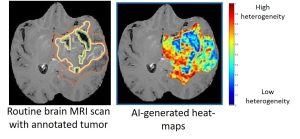
Using what they have learned so far, the researchers plan to conduct a pilot clinical trial that will use their imaging technology to help surgeons identify the best location to biopsy. Comparing the biopsy findings with their computational approach will demonstrate how well their technology is able to distinguish between regions of radiation effects and tumor recurrence.

“We’ll create brain maps that the surgeon can use to navigate to the best location from which to take a biopsy,” said Tiwari. “This will be one of the first clinical trials to use machine learning for biopsy navigation in brain tumors.”

Tiwari says that obtaining funding for this type of pilot study can be difficult because it is a “high-risk, high-reward” project. If successful, it will demonstrate that the new method works well enough to be integrated in larger clinical trials while also giving clinicians more confidence in using machine learning and artificial intelligence approaches. Findings from the V Foundation project have also helped Tiwari secure an NIH grant that will support additional development of these tools.

Canine companions could bring new insights into human cancer

Canine companions could bring new insights into human cancer

Approximately 80,000 Americans are diagnosed with bladder cancer each year. Although new immunotherapies known as immune checkpoint inhibitors are very promising, they are only effective in about 25% of patients with this cancer. Despite numerous studies, there is still no clear understanding of why the majority of patients with bladder cancer do not respond, nor are there clear ways to predict which patients will respond. Understanding the mechanisms of resistance to checkpoint inhibition has been frustrating and expensive, leaving both patients and doctors wishing for a better way.

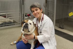
With support from the V Foundation, Nicola J. Mason, B.Vet.Med., Ph.D., at the University of Pennsylvania School of Veterinary Medicine, is trying to find a better way to match checkpoint therapies with patients most likely to respond to them by studying bladder cancer in dogs. Her goal is twofold: to provide new treatments for dogs suffering from bladder cancer and to discover new biomarkers to identify which human and canine patients are most likely to benefit from these immunotherapies.

A win-win approach to cancer research

Mason’s project is funded through the V Foundation’s canine comparative oncology grant program, which supports researchers from human and veterinary medicine using a comparative approach that could lead to better cancer therapies for humans and pet dogs.

For years, mice have been used to study cancer. Although many important discoveries have been made using mice, they are model systems and frequently don’t show the same types of side effects that people experience in response to cancer therapies. Pet dogs are 85% similar to humans genetically and develop spontaneous tumors, including bladder cancer, that share many biological and behavioral characteristics with human cancer. Furthermore, pet dogs have intact immune systems and are likely to be highly valuable in determining responses to immunotherapies.

It was insightful and visionary for the V Foundation to recognize the shortfalls of studying cancer and immunotherapies in mice and to address these issues head-on by building a program around canine comparative oncology. This approach creates a win-win situation, as through it we learn information that could be used to treat cancer more effectively in people while also potentially improving the lives of our companion animals.
Nicola J. Mason, DVM, Ph.D.

Developing state-of-the-art tools to study canine cancer

Bladder cancer in dogs is surprisingly similar to bladder cancer in people. These tumors tend to have a similar genetic makeup, the same subtypes and similar responses to chemotherapy. Mason is leading an interdisciplinary team of researchers from the University of Pennsylvania Veterinary and Medical schools to examine the biology of canine bladder cancer from several different angles.

“We are developing advanced tools for studying bladder cancer in dogs,” said Mason. “Having immune reagents that parallel those used to treat and assess response to treatment in people should help us address this question of why some human bladder cancer patients respond to immunotherapy while others do not. Eventually we may be able to use this information to help more patients respond to this treatment.”

They are specifically looking at checkpoint inhibitors, which are designed to unleash the full power of T-cells to fight cancer by blocking certain immune checkpoints that would normally keep the body’s T-cells from overreacting during an immune response. The researchers have developed their own checkpoint inhibitors for use in dogs because canine versions are not yet available for clinical use. So far, they have developed the canine equivalents of two human checkpoint inhibitors, which they plan to test soon. Before treatment, they will take biopsies of bladder tumors that have developed spontaneously in pet dogs to analyze genetic mutations and the types of cells present in the tumor microenvironment. After treatment with the checkpoint inhibitor, they will take another biopsy to see how the tumor changes in response to the therapy.

“We predict that the response to checkpoint inhibition will positively correlate with tumor mutational burden, and it may also be associated with the baseline immune profile of the tumor,” Mason said.

For quick and inexpensive analysis of a tumor’s mutational burden, they have developed a next-generation sequencing panel that can identify mutations occurring in genes that are commonly mutated in cancer. They are currently validating this tool, which corresponds to panels used to study tumor cell mutations in people.

In another aim of this study, the researchers are examining biomarkers that could be used to predict cancer response or resistance to checkpoint inhibitors. For this, they worked with the life science company NanoString to design a gene expression panel that can be used to evaluate the genes that make up the immune landscape (immunome) of cancer samples. The panel closely parallels one designed to evaluate the immunome in human cancer samples. The canine panel allows the researchers to examine 800 genes covering about 47 different immunological pathways and will be employed to analyze gene expression before and after checkpoint inhibitor treatment. They are also developing immunofluorescent strategies to determine the types of immune cells that infiltrate the tumors after checkpoint inhibition.

Looking beyond bladder cancer

The tools the researchers are developing with V Foundation funding can also be used to study other types of cancer. For example, the researchers are beginning to look at the mutational burden of hemangiosarcoma, a deadly cancer in dogs that is similar to a rare cancer in people called angiosarcoma. Although it is a very serious cancer, angiosarcoma is extremely difficult to study in people because there are so few patients. The researchers also plan to examine immune responses to checkpoint inhibition in other cancers and determine if clinical response correlates with tumor mutational burden and baseline immune status of the tumor.

“In these cancers, it is possible that we will find that the mutational load and immune response to checkpoint inhibition in dogs and humans are similar,” said Mason. “This will open up many new avenues for using the dog as a model to understand responses to checkpoint inhibitors and provide insights into basic tumor biology and immunology. This will benefit both the dogs and people.”

Understanding cancer’s chaotic chromosomes

Understanding cancer’s chaotic chromosomes

Peer through a microscope at almost any cell in your body and you’ll find 23 pairs of chromosomes—neatly coiled bundles of DNA containing all of the genetic instructions needed to make your body and keep it running. But look at a cancer cell and you may notice something unusual. Instead of the typical 23 pairs, many tumor cells have extra or missing chromosomes, a condition called aneuploidy. Solving the mystery of why cancers exhibit aneuploidy could help scientists develop more effective treatments, including therapies that can better target cancer cells while sparing normal cells.

However, studying aneuploidy isn’t easy. Scientists can inactivate single genes to identify their function, but the addition of just one chromosome affects the expression of thousands of genes. In an important advance aided by a V Scholar grant, Teresa Davoli, Ph.D., from the New York University School of Medicine is developing a new technology that makes it possible to add or delete individual chromosomes in cells.

“Much of today’s cancer treatment research is focused on specific genetic mutations that are usually present in a relatively low number of patients,” said Davoli. “Because aneuploidy is present in virtually all patients with solid tumors, understanding which cells are vulnerable and the role aneuploidy plays in cancer could lead to treatments that help a lot of people.”

A tool for chromosome editing

The ability to create aneuploidy in the lab can help scientists study how this condition arises in cancer cells and potentially inform cancer treatments. Davoli and her team plan to use the new method to insert extra chromosomes into normal cells and also fix aneuploidy in cancer cells by removing the extra copies.

“To learn more about what aneuploidy is doing in tumor cells, we can compare cells in which we’ve added chromosomes to those with which we’ve removed chromosomes,” said Davoli. “One of the first cancers we want to examine is colorectal cancer, which tends to exhibit extra copies of chromosome 7 or 13.”

Chromosome-specific aneuploidy might also prove to be a useful biomarker, or indicator, of how a patient will respond to certain therapies. “Work we’re conducting in collaboration with Scott Lippman, Director of the Moores Cancer Center at the University of California San Diego, is showing that the deletion of specific chromosomes was associated with a poor response to immunotherapy in head and neck cancer,” said Davoli.

Applications beyond cancer

Davoli is also collaborating with New York University researchers who want to use the new technology to study other diseases that involve aneuploidy – such as Down syndrome. It might also be useful for examining how certain biological processes differ between men and women.

Davoli says that the V Foundation funding was crucial to giving her the time to develop the new technique, a process that has taken several years. “Our proposal was relatively risky, and the aneuploidy field is more obscure than other areas of cancer research,” she said.

The work we did with V Foundation support has led to early-stage investigator funding from the National Institutes of Health to continue with this research.
Teresa Davoli, PH.D.


Eavesdropping on Cancer-Killing Cells, with Help from Ancient Bacteria

Eavesdropping on Cancer-Killing Cells, with Help from Ancient Bacteria

In the past few decades, immunotherapies have emerged as an important treatment option for many cancers. These treatments activate our immune system, priming it to recognize and destroy cancer cells. While these therapies offer great promise, there’s still much work to do. Scientists are still trying to understand why some patients don’t respond to these treatments and why new immunotherapies that work well in animal models don’t always translate to people.

With support from the V Foundation, Philip Kranzusch, PhD, from the Dana-Farber Cancer Institute is studying how immune cells communicate with each other to fight cancer. He said he hopes decoding the signals cells sent through a pathway known as cyclic GMP–AMP synthase (cGAS) stimulator of interferon genes (STING) could help scientists optimize immunotherapies that involve these signals.

“cGAS-STING is a signaling pathway that allows immune cells to sense foreign DNA, including that from tumors,” said Kranzusch. “Although we know that STING is activated in immune cell responses, many aspects of how it works remain unknown.”

Looking to bacteria for answers

Although quite a few researchers have studied the human cGAS-STING pathway, Kranzusch’s team decided to take a different approach by looking to ancestrally related proteins to tease apart details of how this pathway works.

“The human version of STING can be thought of as a complex machine with multiple layers of regulation,” said Kranzusch. “Looking for differences between STING found in primitive systems and humans can tell us about its function and regulation in people.”

In work led by Benjamin Morehouse, PhD, a senior postdoctoral fellow in Kranzusch’s lab, the researchers made the surprising discovery that STING also exists in bacteria. Building on this finding, they compared the molecular structure of bacterial STING proteins with that of human STING proteins.

“We discovered that bacterial STING proteins are much simpler than what is found in humans,” said Morehouse. “This let us identify the core machine function and then see how layers of regulation have been added on in humans, adding more clarity to the mechanisms of how STING functions.”

This work, published in Nature, shows cGAS-STING is much more ancient than scientists thought. It also offers a new approach that can be used to answer more questions about how these proteins actually work in human cells.

“We think that hundreds of millions of years ago, this pathway originated in bacteria as a way to defend themselves against viruses,” said Kranzusch. “Then through evolution, it was transferred into animal genomes and eventually became the human pathway that’s important for cancer immunotherapy.”

Improving treatments

The team’s discoveries could help inform cancer treatments that target STING, some of which are in development at pharmaceutical companies. Armed with new knowledge about STING structures and which pieces are unique to humans, scientists can design medicines capable of generating a stronger antitumor response in more patients.

Kranzusch credits V Foundation donors with kick-starting this fruitful vein of discovery.

The V Foundation’s support for the lab has been incredible,” he said. “All of the many projects that have grown out of this research have been because the V Foundation was one of the first to take the chance with our lab and support our research.
Philip Kranzusch


Making Gynecologic Cancer Screening Simpler, Safer with At-Home Test Kit

Making Gynecologic Cancer Screening Simpler, Safer with At-Home Test Kit

The number of women who develop cervical cancer has been greatly reduced due to screening with Pap smears, but other gynecologic cancers such as ovarian and endometrial cancer, remain difficult to detect at an early stage, when treatments are more effective.

This year alone, over 65,000 new cases of endometrial cancer will be diagnosed in the U.S. Sadly, for many women, the diagnosis will come too late, when the cancer is already advanced. Jamie Bakkum-Gamez, M.D., a gynecologic oncologist from the Mayo Clinic, hopes to turn the tables on this potentially devastating disease by developing the first early detection diagnostic test for endometrial cancer.

Bakkum-Gamez and her team are working to create a simple test kit that women could use in the privacy of their own home to screen for endometrial, ovarian and cervical cancer all at once. Women would use the kit to collect a sample of vaginal fluid and then send it to a lab for analysis.

“The pandemic has underscored the importance of remote patient care and telehealth as tools that can help people stay as healthy as possible,” Bakkum-Gamez said. “Tests like the one we are developing are another important component of this because they can be done at home without risking exposure to COVID-19 by going into a public space like a doctor’s office.”

Searching for cancer’s telltale signs

The team’s test is designed to detect the epigenetic DNA modification known as methylation, which changes the way a gene is expressed without changing the gene’s DNA sequence. When looked at collectively, these modifications across all DNA within the cells of each unique tissue in the body are referred to as the methylome. In a pilot study, the researchers showed markers based on DNA methylation unique to endometrial cancer could be detected in the vaginal fluid of women with endometrial cancer but are not present in vaginal fluid of women without cancer.

Thanks to V Foundation support, Bakkum-Gamez then formed a collaboration with the team at Mayo Clinic that developed Cologuard, an at-home test used for colon cancer screening. Cologuard makes it simple to collect a stool sample at home and then ship it to a lab, where it is analyzed for colon cancer biomarkers.

“With the same technology used to develop Cologuard, we performed an extremely thorough sequencing-based search through the DNA methylome to find biomarkers that were associated with endometrial cancers, as well as ovarian and cervical cancers,” said Bakkum-Gamez. “We are the first group to define the methylome for endometrial cancers.”

Bakkum-Gamez and her team identified unique markers for the most common form of endometrial cancer, which tends to be the easiest to treat, as well as forms of endometrial cancer that are more aggressive. To see whether these biomarkers could be used to identify cancer using vaginal fluid, they conducted a clinical trial involving 200 women, half of whom had endometrial cancer and half of whom had benign gynecologic conditions. Participants collected their own vaginal fluid by inserting and later removing a regular over-the-counter tampon.

“Using a panel of 29 methylation markers, we were able to identify a very high proportion of the women with endometrial cancer just based on vaginal fluid collection and testing,” said Bakkum-Gamez. “Because of these promising findings, we are planning a larger study with a more diverse group of participants.”

Beyond endometrial cancer

The researchers have also identified unique methylation markers for other gynecological cancers. These markers will also be tested in vaginal fluid. “Because there may be different markers for different types of cancers in the vaginal fluid, we wanted to make sure that we weren’t missing an opportunity to detect other cancers,” Bakkum-Gamez said. These markers eventually could be incorporated into an at-home test that screens for endometrial, cervical and ovarian cancers.

If the larger clinical studies continue to be successful, Bakkum-Gamez estimates an at-home screening test could potentially be available in the next five years.

The V Foundation support helped our research move forward at a much faster rate than would otherwise be possible. It also enabled our collaboration with colleagues that developed the Cologuard test, which has now led to an industry partnership with Exact Sciences that will help translate this technology into a useful test for patients.
Jamie Bakkum-Gamez

An easy-to-use, at-home gynecologic cancer test kit would not only be useful to patients—it would also bring peace of mind. With the promising capability to detect gynecological cancers earlier than ever before, Bakkum-Gamez hopes women will soon be able to take comfort in knowing that if cancer does strike, it can be caught in time for lifesaving treatment.

Refusing to Give Up on Older Patients

Refusing to Give Up on Older Patients

Acute myeloid leukemia (AML) is the most common form of acute leukemia in adults, with more than 20,000 new cases diagnosed in the U.S. every year. One of the biggest mysteries of this disease is how differently it behaves in patients of different ages. When it strikes before age 25, AML is very treatable, and the vast majority of younger patients can expect to live long, full lives. For patients over 60, however, the outlook is dramatically worse: The 5-year survival rate is less than 20%, and deadly relapses are much more common.

Francine Garrett-Bakelman, M.D., Ph.D., of the University of Virginia School of Medicine, has been determined to change those outcomes ever since she first encountered AML during her hematology and oncology clinical training rotation at Weill Cornell Medicine as a medicine resident. Its effect on older patients was particularly fascinating, and frustrating, to see.

“I was humbled by the complexity of the disease and by how much patients suffer from it,” she said. “It’s one of those situations in medicine where you can really make a difference by extending survival by months or years, and helping someone through a very challenging time and treatment.”

New insights for older patients

In order to help this group of vulnerable patients live longer, Garrett-Bakelman and her team set out to decipher the role of epigenetics—changes in the expression of genes—in AML. Although our genes rarely change as we age, changes affecting the expression of genes accumulate over our lifetime and influence our health. With cancer, the relationship goes both ways: Epigenetic changes influence cancer development and the response to treatment, and cancer and its treatments can also cause epigenetic changes.

Using tissue samples from a group of older AML patients, Garrett-Bakelman and her team analyzed the expression of two genes they detected in AML: RBM47 and FBP1. They found higher expression of these genes was associated with better survival rates, even in this high-risk group.

The findings point to a possible explanation as to why some treatments work better in younger patients than older ones. At a minimum, this information may help doctors determine which older patients are most likely to benefit from available treatments. Better yet, researchers could search for new treatments that target those genes differently and may be more effective in older people.

Our goal is to use this precious material in a way that can teach us why older people developed the disease and what the biomarkers are telling us. Can they determine who should get treatment and how?
Francine Garrett-Bakelman, M.D., Ph.D.

In related follow-up studies, the team is also investigating if differences in gene expression—and the resulting differences in outcomes—could be influenced by epigenetic changes that are themselves caused by cancer therapies.

Starting out strong

Garrett-Bakelman, whose 2017 V Scholar Grant was funded by the Stuart Scott Memorial Cancer Research Fund, has been supported by the V Foundation from the start of her career, and she sees it as integral to her work. In addition to her current V Foundation grant, Garrett-Bakelman was also a Virginia Vine Team investigator in 2019.

“The funding the V Foundation offers is invaluable, especially to people like myself who are just starting out in their careers,” she said. “It has helped me focus on the science and generate exciting results I can share with others, as we strive to make progress in understanding AML.”

The future for AML

Garrett-Bakelman and her team are continuing to investigate whether epigenetic mechanisms are in fact controlling gene expression in high risk AML patients. Because of the complex interplay between genes and cancer, an answer won’t come overnight, but she is encouraged by the team’s early results.

With enough time and study, Garrett-Bakelman is confident the important questions can be answered, and eventually, patients of any age will be able to look forward to many healthy years ahead after what is, for now, a devastating diagnosis.

Intercepting secret messages to fight pancreatic cancer

Intercepting secret messages to fight pancreatic cancer

Researchers have worked tirelessly for decades to find new therapies for pancreatic cancer, yet it remains a difficult disease to treat. In fact, in 2016, pancreatic replaced breast cancer as the third leading cause of cancer-related deaths in the U.S.

“There have been significant strides in understanding the genetics of pancreatic cancer,” said Mara Sherman, Ph.D., from the Oregon Health & Science University Knight Cancer Institute. “However, it is becoming clear that we need to know more about the biology of this cancer to develop more effective treatments.”

With V Foundation funding, Sherman is moving science closer to understanding the biological pathways that make pancreatic tumors tick. Among cancers, one thing that sets pancreatic tumors apart is their unusual microenvironment. Only a small portion of the tumor consists of cancer cells—malignant cells with mutated DNA. Most of the tumor is made of scar-like tissue that nurtures the cancer cells while shielding them from anti-cancer drugs. Sherman said she believes understanding the inner workings of this protective tissue could be key to developing new therapies for pancreatic cancer.

Looking to the stars

Sherman’s research focuses on stellate cells, which are shaped like tiny stars. In healthy tissue, these cells help to heal damage in the pancreas, liver and other organs by transforming themselves into scar-forming cells called myofibroblasts. They also are the body’s storage lockers for vitamin A, which they carry inside small droplets of fat, or lipid.

Stellate cells respond to a tumor the same way they respond to tissue damage, by creating scar tissue. Unfortunately, in the case of pancreatic cancer, it seems this process may only make the situation worse.

My previous work showed that lipid storage and metabolism change profoundly in stellate cells during pancreatic tumor progression. In this new work, we wanted to understand the potential significance of this and whether this switch may somehow help pancreatic cells survive.
Mara Sherman, Ph.D.

Working with Jurre Kamphorst from the Beatson Institute in the U.K., Sherman and her team found fibroblasts from human pancreatic tumors secrete high levels of lipids. Using mouse models, they also observed pancreatic cancer cells take up these lipids. These findings suggest stellate cells are essentially feeding the cancer by spitting out fat droplets.

Following the trail further, the researchers discovered the lipids are also metabolized, or digested, by autotaxin, an enzyme secreted from pancreatic cancer cells. This process creates a molecular signal that urges the cancer cells to keep growing.

While it would likely be hard to stop stellate cells from feeding their lipids to cancer cells, stopping autotaxin is much more feasible. In fact, there are already drugs on the market that inhibit autotaxin. To find out how inhibiting autotaxin would affect the cancer, the researchers blocked the enzyme using two different methods: drugs and genetic manipulation. Although both approaches slowed pancreatic tumor progression in mice, the genetic inhibition was much more potent. This could mean the drug wasn’t getting delivered efficiently, or that a better drug is needed.

Targeting the ‘puppet master’

The researchers are continuing to study how inhibiting autotaxin affects pancreatic cancer cells and the tumor microenvironment. “If we better understand the consequences of autotaxin inhibition, perhaps we can come up with a combination of therapeutic strategies that will be more efficient than targeting autotaxin alone,” said Sherman. “Understanding the cell type that’s the puppet master of this reaction may help us figure out which pathways or molecules to target therapeutically.”

To do this, the researchers are developing new mouse models that will allow them to turn off specific cell types, enabling them to see what happens to other cells within the tumor microenvironment when certain cells are inactivated.

“When we applied for the V Foundation funding, we didn’t know if the process we were studying would actually impact tumor progression,” said Sherman. “The support allowed us to get preliminary data, which led to an NIH grant that is allowing us to continue this line of study.”

Sherman said she hopes that within the next five to 10 years their research will reveal a combination of drugs that is promising enough to move into clinical testing.

In Their Own Words: Trea Turner

In Their Own Words: Trea Turner

The V Foundation for Cancer Research is successful thanks to the contributions of many – donors, corporate partners, our incredible Board and Scientific Advisory Committee and the amazing researchers to whom we award grants. With “In Their Own Words,” we sit down with key members of our team to learn more about their commitment to the V Foundation and their personal desire to put an end to cancer. In this edition, we chat with World Series champion Washington Nationals shortstop Trea Turner.


The V Foundation: How has cancer affected you personally?

Trea Turner: Cancer has affected both of our families. Kristen and I have seen loved ones battle with cancer, and we know the feelings of helplessness and frustration it brings to everyone involved. We have been moved by our own experiences to help the efforts to find a cure to this terrible disease.


The V Foundation: What made you decide that you wanted to support pediatric cancer research specifically?

Trea Turner: Kristen and I attended NC State University, where the legendary Jim Valvano coached. We knew of his story and the difference he made in the battle with cancer. We have wanted to give back and make a difference as well. We love kids and thought we could make the biggest impact in support of the fight against pediatric cancer.


The V Foundation: How has Jim’s legacy inspired you (and other members of the NC State community) to support cancer research?

Trea Turner: He is a huge part of the reason we got involved. He is a great example of perseverance and positivity. We loved his message of never giving up, and we feel a strong connection with him through the NC State community.


The V Foundation: You encouraged fans to join in on your efforts in raising money for research via Pledge It last season. Why was it so important for you to give fans the chance to join you in supporting research?

Trea Turner: I think when you can have a group of people get behind a great cause and all pull in the same direction great things can happen. One person can make an incredible difference, but with the addition of each individual, the possibilities are endless. We wanted to give the pediatric cancer community a cause to get behind so that we are in this together, just like sharing in the journey of a baseball season.


The V Foundation: How has your life changed after winning the World Series in 2019?

Trea Turner: Honestly, my life hasn’t changed much. Other than the fact that I have a beautiful World Series ring that I’ll always love, along with the greatest memories with an unbelievable group of guys that I’ll never forget!


The V Foundation: What would Victory Over Cancer® look like to you?

Trea Turner: Victory over cancer to me would look like freedom – free from suffering, free from pain, free from seeing someone with so much life in front of them gone.

Posts navigation

Newer posts
Follow Us
Language
Get In Touch

The V Foundation for Cancer Research, 14600 Weston Parkway, Cary, NC 27513
Email: [email protected]    Phone: 919-380-9505

Charity Navigator logo Candid GuideStar logo
About Us
  • Our Mission
  • Financial Responsibility
  • Leadership
  • Partners
  • Ambassadors
  • Careers
Events
  • Signature Events
Research & Impact
  • Research
  • Key Research Initiatives
  • Proven Positive Impact
  • Award Programs
  • Grant Process
  • Funded Grants
Other
  • Newsroom
  • Stories
Take Action
  • Ways to Give
  • DIY Fundraising
  • Run For Team V
  • Cause Marketing
Copyright © 2026 V Foundation for Cancer Research. All rights reserved.
The V Foundation for Cancer Research is a 501(c)(3) charitable organization. Federal Tax ID Number is 13-3705951.
Legal Privacy Policy
Manage Consent
To provide the best experiences, we use technologies like cookies to store and/or access device information. Consenting to these technologies will allow us to process data such as browsing behavior or unique IDs on this site. Not consenting or withdrawing consent, may adversely affect certain features and functions.
Functional Always active
The technical storage or access is strictly necessary for the legitimate purpose of enabling the use of a specific service explicitly requested by the subscriber or user, or for the sole purpose of carrying out the transmission of a communication over an electronic communications network.
Preferences
The technical storage or access is necessary for the legitimate purpose of storing preferences that are not requested by the subscriber or user.
Statistics
The technical storage or access that is used exclusively for statistical purposes. The technical storage or access that is used exclusively for anonymous statistical purposes. Without a subpoena, voluntary compliance on the part of your Internet Service Provider, or additional records from a third party, information stored or retrieved for this purpose alone cannot usually be used to identify you.
Marketing
The technical storage or access is required to create user profiles to send advertising, or to track the user on a website or across several websites for similar marketing purposes.
  • Manage options
  • Manage services
  • Manage {vendor_count} vendors
  • Read more about these purposes
View preferences
  • {title}
  • {title}
  • {title}
Manage Consent
To provide the best experiences, we use technologies like cookies to store and/or access device information. Consenting to these technologies will allow us to process data such as browsing behavior or unique IDs on this site. Not consenting or withdrawing consent, may adversely affect certain features and functions.
Functional Always active
The technical storage or access is strictly necessary for the legitimate purpose of enabling the use of a specific service explicitly requested by the subscriber or user, or for the sole purpose of carrying out the transmission of a communication over an electronic communications network.
Preferences
The technical storage or access is necessary for the legitimate purpose of storing preferences that are not requested by the subscriber or user.
Statistics
The technical storage or access that is used exclusively for statistical purposes. The technical storage or access that is used exclusively for anonymous statistical purposes. Without a subpoena, voluntary compliance on the part of your Internet Service Provider, or additional records from a third party, information stored or retrieved for this purpose alone cannot usually be used to identify you.
Marketing
The technical storage or access is required to create user profiles to send advertising, or to track the user on a website or across several websites for similar marketing purposes.
  • Manage options
  • Manage services
  • Manage {vendor_count} vendors
  • Read more about these purposes
View preferences
  • {title}
  • {title}
  • {title}