Susanna’s Cool Cancer Research Finds: April 2024
Every week, Dr. Susanna Greer, the V Foundation's Chief Scientific Officer, shares her cool cancer research finds on LinkedIn. Read more below to see highlights from the latest top finds!
March 28, 2024
A sophisticated strategy providing cellular insights to treatment resistance in Glioblastoma.
In this week’s Cool Cancer Find, the V Foundation grantee Dr. Sandro Matosevic and team at Purdue University present findings representing a significant breakthrough in cancer research, particularly in our understanding and treating glioblastoma, or GBM, a highly aggressive brain tumor.
Dr. Matosevic and team focused on understanding how certain proteins, particularly those with Immunoglobulin domains, (which are like specialized tools used by the immune system to recognize and neutralize threats, akin to keys fitting into locks), and ITIM domains, (which act as switches to regulate immune responses, similar to how skilled security personnel maintain a balanced approach in handling situations), interact with each other within cells. These interactions play a crucial role in modulating immune responses.
 The researchers identified two key proteins in glioblastoma contributing to the tumor’s resistance to treatment: the T cell immunoreceptor with Ig and ITIM domains (or TIGIT) and CD73, both of which play critical roles in suppressing the immune system’s ability to fight cancer.
TIGIT contributes to immune suppression by inhibiting the activity of natural killer cells, which are normally tasked with eliminating cancerous cells. CD73 acts differently by producing adenosine, an immunosuppressive molecule that further hampers the immune response against tumors.
I LOVE this paper and the findings within for two main reasons. First, Dr. Matosevic and team use a sophisticated strategy to combat the immunosuppression: they discovered cellular mechanisms which triggered a cascade of molecular events that not only blocked the immunosuppressive effects, but also released a therapeutic agent. This dual action effectively enhanced the immune system’s ability to recognize and eliminate GBM cells. Incredible!
Second: In preclinical studies, the approach they used showed remarkable efficacy in reducing tumor growth and improving survival rates in mouse models of GBM. Mouse models are one step in a LONG line of developmental, to preclinical to clinical research. But a VERY necessary one, and a difficult hurdle to overcome. The findings in these models are breathtaking.
The findings from this publication represent a significant advancement in cancer immunotherapy in general and particularly for GBM, which has been notoriously difficult to treat. By targeting multiple immunosuppressive pathways simultaneously, this approach holds initial promise for developing more effective and innovative treatments for GBM patients.
Follow the Matosevic lab and their research at Faculty | IMPH – Purdue and read this outstanding paper at synNotch-programmed iPSC-derived NK cells usurp TIGIT and CD73 activities for glioblastoma therapy | Nature Communications
April 5, 2024
Tackling one of cancer immunotherapies biggest successes and biggest challenges, PD-L1, with novel drug design.
This week’s Cool Cancer Find is REALLY COOL. Long time readers, (as in all 4 weeks, ? of my Newsletter + months of LinkedIn posts…thank you for coming along with me and reading!), know I love ALL cancer research, and that I am an Immunologist by training. As such, I have a special soft spot for sharing exciting research finds in cancer immunotherapy. For those new to my posts, or new to cancer immunotherapy in general, let’s back up a bit before we dive in to this week’s Cool Cancer Find:
Today we dip into immunotherapy via an outstanding paper from the V Foundation grantee Dr. Jamie Spangler and her lab at John Hopkins University which is focused on a protein ominously named Programmed Death Ligand 1. What is Programmed Death Ligand 1 you say? Well, well, well…I’m so glad you asked ?…
Programmed Death Ligand 1, or PD-L1, is a protein found on the surface of some cells in our bodies, including cancer cells. PD-L1 plays a crucial role in regulating the immune system’s response to these cells.
Why? How does PD-L1 work? Well, our immune system is like a defense system in our body, always on the lookout for invaders like bacteria, viruses, and, cancer cells. But sometimes, cancer cells can trick the immune system into leaving them alone. They do this by using PD-L1 as a shield.
 PD-L1 acts like a stop sign for our immune cells, telling them not to attack. This helps cancer cells avoid being destroyed by the immune system, allowing them to grow and sometimes spread in our body not cool cancer, not cool.
Understanding PD-L1 has been incredibly important for progress in cancer research. the V Foundation funded researchers and many others in the cancer research community have participated in research leading to the development of drugs called PD-L1 inhibitors that can block the PD-L1 protein. You might have heard of ‘immune checkpoint proteins.’ PD-L1 is the most famous of all immune checkpoint proteins!
PD-L1 is important in cancer research because it’s a target for new treatments that help a lot of people by boosting their immune system’s ability to fight cancer. A significant challenge remains as response rates vary largely across tumors and across patients to PD-L1 inhibitors.
WHEW…so now that we have cliff notes on the incredible discoveries surrounding PD-L1 and how we BLOCK it’s activity with PD-L1 inhibitors, on to this week’s Cool Cancer Find:
Dr. Spangler is an the V Foundation grantee who has made great strides in understanding molecules, specifically proteins like PD-L1, that can be used for therapeutic purposes, particularly in treating diseases like cancer.
 Dr. Spangler and her team use a method called protein engineering, which involves tinkering with proteins to make them do new and useful things. Instead of randomly trying different combinations of proteins and protein ‘parts’, the Spangler lab uses the lab’s growing understanding of how proteins work at a molecular level to guide experiments. Their approach involves using advanced tools and knowledge in areas like structural biology to understand how proteins are shaped, and molecular design to figure out how to change protein structures. By doing this, the researchers can tailor proteins to have specific activities that could be beneficial for treating cancer.
So how does this all relate to PD-L1? Well, the Spangler Lab has taken drug design knowledge to the next step with PD-L1. Having too much PD-L1 (an immune checkpoint) on a tumor has motivated the design of targeted antibodies to disrupt the interaction with PD-1 on immune cells…but remember…despite clinical success of these antibodies, response rates remain low, necessitating novel approaches to enhance performance.
What Dr. Spangler and her team show in this paper is the development of antibody fusion proteins that block immune checkpoint pathways through a new and distinct mechanism targeting molecular trafficking. Their unique method causes PD-L1 to clump together, get pulled inside the cancer cells, and then to break down quickly. This not only boosts the immune system’s ability to fight tumors, but also greatly reduces the amount of PD-L1 on the cancer cells, making them more vulnerable to attack.
These findings are impressive to me for two reasons: first, they show new ways we can manipulate the immune system to improve cancer treatments in the future and second, in animal studies, the cancer cells were left more exposed and vulnerable to attack by the immune system.
Bottom line: the Spangler lab is taking a smart, targeted approach to design new proteins that could become the basis for future drugs against cancer.
Follow the Spangler lab @ Jamie Spangler – Johns Hopkins Biomedical Engineering (jhu.edu) and read her lab’s paper here https://doi.org/10.1016/j.chembiol.2024.02.014
April 19, 2024
An unexpected GOOD sign – tumor associated macrophages and breast cancer.
This week’s Cool Cancer Find highlights a favorite protein in the world of cancer research, and one we’ve talked about in the Newsletter before: programmed cell death ligand 1 (PD-L1). This incredible protein plays a critical role in the immune system’s response to cancer. In my newsletter we often imagine the immune system as an army that fights invaders like cancer cells. PD-L1 is like a signal that some cells, including cancer cells, use to tell the immune system to hold back and not attack.
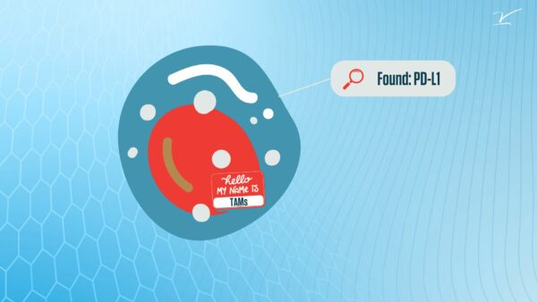 In this really cool study, V Foundation Grantee Dr. Peter Lee at City of Hope wanted to understand how PD-L1 works in a type of immune cell called tumor-associated macrophages or TAMs. TAMs are a kind of immune cell found within tumors. Previous research in the Lee lab had shown that some TAMs have PD-L1 on their surface, while others don’t. Dr. Lee and team wondered what this difference might mean for how TAMs interact with the immune system and cancer cells.
To answer that question, Dr. Lee investigated the genetic makeup of these TAMs in breast cancer patients. They found that TAMs with PD-L1 seemed more active compared to those without PD-L1. These PD-L1 positive TAMs also seemed to be better at stimulating other immune cells called T cells, which are important for fighting cancer.
Surprisingly, they discovered that breast cancer patients with more PD-L1 positive TAMs tend to have better outcomes. This finding was counter intuitive because PD-L1 usually tells the immune system to hold back. But in this case, it seemed like PD-L1 positive TAMs might actually help the immune system fight cancer.
To understand this better, the Lee team looked at how these different TAMs interact with other cells in the tumor. They found that PD-L1 positive TAMs tend to interact more with T cells, while PD-L1 negative TAMs interact more with cancer cells. Their findings suggest that PD-L1 positive TAMs might be helping to recruit T cells to fight the cancer. Very cool, right?
 I love this study because it helps shed light on how different types of immune cells in tumors can affect cancer progression in different ways. It also reminds ME to keep an open mind about cancer immunotherapy as it suggests that PD-L1 positive TAMs might be a good sign in breast cancer patients, as they could be helping the immune system fight the cancer cells.
Follow the Lee lab and their research at: Peter P. Lee, M.D. | City of Hope and read their paper here: PD-L1-expressing tumor-associated macrophages are immunostimulatory and associate with good clinical outcome in human breast cancer: Cell Reports Medicine